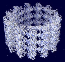
Space-filling model of a microtubule segment derived from cryo-electron microscopy. The protofilaments are seen running along the axis of the segment. The microtubule (+) end is towards the top of the image. (Source: Wikimedia commons)
Episode 12 - The Cytoskeleton
The cytoskeleton functions to maintain cell organization, shape, and to allow transport within the cell to occur. There are three main types of cytoskeleton which are discussed below.
Microtubules
Microtubules are the largest physical components of the cytoskeleton. The microtubule is mainly made up of two types of ball-shaped protein monomers (α- and β-tubulin), which bond together into tubulin dimers.
Microtubules maintain cell shape: the ones that grow out from centrosomes near the nucleus (the center) of the cell act as compression-resistant girders for the cell, preventing the cell from being squashed. Microtubules also interact with motor proteins: Motor proteins (especially Kinesin) that attach to motor-protein-receptors on various organelles (like vesicles) can carry the organelles on microtubule rail-tracks around the cell (with ATP as energy input).
Space-filling model of a microtubule segment derived from cryo-electron microscopy. The protofilaments are seen running along the axis of the segment. The microtubule (+) end is towards the top of the image. (Source: Wikimedia commons)
Microtubules also exist in cilia and flagella in a “9+2” array and thus contributes to cell motility. Microtubules are also responsible for pulling apart duplicated chromosomes during cell division:
Microtubules are very important to the cell, and are thus much targeted by poisons such as colchicines and taxol, which either disassembles existing microtubules or prevent the formation of new microtubules.
Actin Filaments
Microfilaments, also called Actin Filaments, are the smallest of the three types of “cytoskeleton parts”. They are solid rods that are present in all eukaryotes. Microfilaments are made up of two polypeptide chains, assembled from actin monomers, twisted in a helix shape between 5-7 nm in diameter. Certain proteins can link adjacent microfilaments together, and microfilaments can form branches with itself. This boning pattern creates a three-dimensional network of proteins and microfilaments beneath the cell membrane, which reinforces cell shape and resists tension (pulling) forces on the cell.

Actin cytoskeleton of mouse embryo fibroblasts, stained with Fluorescein isothiocyanate-phalloidin. (Source: Wikimedia Commons)
Projections of microfilaments make up the core of microvilli. Also gives the cortex (the outer cytoplasmic layer of the cell) a gel-like consistency (less fluid).
Microfilaments, along with myosin filaments, function in muscle contractions. (Myosin acts like a motor-protein, with little arms that walk along the actin filaments, which result in muscle contraction.) Actin-myosin aggregates are also responsible for localized cellular contractions:
Actin-myosin interactions are also believed to coordinate cytoplasmic streaming found in large plant cells: the inner, fluid cytosol (the sol) circles around the cell along a bed of microfilaments parallel to the direction of cytosol flow. Myosin attached to organelles in the sol interacts with the actin to move through the cytosol.
Intermediate Filaments
Intermediate filaments are made up of four protofilaments (with varying proteins) twisted together, and are usually between 8-12 nm in diameter. There are six groups of intermediate filaments, based upon the type of protein that makes them up. These are: keratins; desmins and vimentins, which help muscle cells contract; lamins; neurofilaments; and glial fibrillary acidic proteins.

Structure of intermediate filament. (Source: Wikimedia Commons)
Intermediate filaments function to strengthen cells and cell elements and help maintain their shape. Like microfilaments, intermediate filaments are also tension-bearing.
Intermediate filaments are the most stable component of the cytoskeleton. Thus they are very important in maintaining cell shape and fixing the positions of organelles within the cell.
Intermediate filaments also participate in desmosomes, or anchoring junctions, and help anchor the junction into the cytoplasm of both cells involved in the junction. Each cell has only about two types of intermediate filaments. Thus, biologists can use the type of intermediate filament found in a cell to determine cell type.